𝗕𝗮𝗯𝗲𝘀𝗶𝗼𝘀𝗶𝘀 (𝗥𝗲𝗱 𝗪𝗮𝘁𝗲𝗿)
𝑰𝒏𝒕𝒓𝒐𝒅𝒖𝒄𝒕𝒊𝒐𝒏
⇨ Babesiosis is a protozoal tick-born disease characterized by extensive intravascular hemolysis leading to depression, anemia, icterus, hemoglobinuria, and in the case of B. bovis infections neurological signs
⇨ It is OIE listed disease
⇨ Babesiosis is a protozoal tick-born disease characterized by extensive intravascular hemolysis leading to depression, anemia, icterus, hemoglobinuria, and in the case of B. bovis infections neurological signs
⇨ It is OIE listed disease
 |
| Babesiosis |
𝗦𝘆𝗻𝗼𝗻𝘆𝗺𝘀
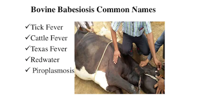
⇨ Redwater
⇨ Tick Fever
⇨ Cattle Fever
⇨ Texas Fever
⇨ Piroplasmosis
𝗘𝗧𝗜𝗢𝗟𝗢𝗚𝗬
⇨ Order:- Piroplasmida
⇨ Family:- Babesiidae
⇨ Genus:- Babesia
⇨ Babesia organisms are typically classified as large (2 to 4 μm) or small (<2 μm) with routine light microscopy
⇨ Babesia spp. are typically host specific and over 100 Babesia species have been identified
⇨ Cattle: Babesia bigemina (large), Babesia bovis (small)
Few reports of B. bovis infection in India
⇨ Horses: Babesia caballi (large)
⇨ Sheep and goats: Babesia motasi (large), Babesia ovis (small)
⇨ Dogs: Babesia canis (large), Babesia conradae, B. gibsoni (small)
⇨ Cats—Babesia cati, Babesia felis, Babesia herpailuri (small)
⇨ Wildlife: B.odocoielei
⇨ Porcine: B. trautmanni and B. perroncitoi
⇨ Babesia case reported in human B. divergens in France, Britain, Ireland, Spain, Sweden, Switzerland and B. microti infections have also been in Taiwan, Japan, and Europe.
Babesia (WHITE color) & Theileria (RED color) mixed infection
𝗛𝗢𝗦𝗧
⇨ Cattle and buffaloes
⇨ Sheep and goats
⇨ Horse; Dog; Cat
⇨ Calves have a degree of immunity that persists for 6 month
⇨ Animals that recover from Babesia infections are generally immune for their commercial life (4 yr),
⇨ Bos indicus cattle tend to be more resistant to ticks and the effects of B bovis and B bigemina infection than Bos taurus–derived breeds
𝗧𝗥𝗔𝗡𝗦𝗠𝗜𝗦𝗦𝗜𝗢𝗡
⇨ Transmitted by ticks
⇨ Babesia bigemina, Babesia bovis Rhipicephalus microplus and R. annulatus also known as Boophilus ticks.
⇨ In ticks, transovarial transmission occurs
⇨ Other routes of minor importance
⇨ Direct inoculation of blood
⇨ Biting flies and contaminated fomites
⇨ In utero infection (rare event).
𝗣𝗮𝘁𝗵𝗼𝗴𝗲𝗻𝗲𝘀𝗶𝘀
Sexual phase within the tick’s gastrointestinal tract
⇓
schizogony resulting in large motile vermicules and this vermicules migrate to tissues, especially the ovary and invade the eggs (transovarial transmission)
⇓
vermicules continue to multiply within the eggs and larval tissues
⇓
When the larval tick moults into the nymph stage, the parasites enter the salivary gland and undergo a series of binary fissions
⇓
They multiply further until the host cells are filled with thousands of minute parasites
⇓
These become infective sporozoites, break out of the host cell, lie in the lumen of the gland, and are injected into the host when the tick feeds
⇓
sporozoites enter into RBCs and within RBCs parasite divide asexually into 2 or 4 pear shaped merozoites (“piroplasm”)
⇓
Merozoites come out from RBCs by lysis of RBCs (intravascular hemolysis)
⇓
Infecting new erythrocytes and many such cycle leads to marked destruction of RBCs (Hemolysis due to direct damage to erythrocytes by protozoal proteases, immune-mediated destruction, or oxidative damage)
⇓
anemia and hemoglobinemia (Increased Hb in plasma/blood) → Hemoglobinuric nephrosis (hemoglobinuria) → Severe anemia
⇓
Parasite is the source of proteases that activate plasma kallikrein (hypotensive agent), may activate bradykinin vasodilatory agent)
⇓
so RBCs are sequestered in capillaries rather than large veins (congestion)→ also elevate anemia
⇓
Parasite proteases hydrolysed fibrinogen so accumulation of large quantities of soluble fibrin in the circulation and increased coagulability and viscosity of blood → extensive plugging of microvasculature by sequestered RBCs
⇓
The combination of vascular congestion, vasodilation, and hemolysis leads to both metabolic alkalosis (B. bovis) and a hemodynamic crisis, hypoxic cell damage
⇓
Irreversible shock → death
⇨ In B. bovis infection, RBCs are sequestered in capillaries of brain → blocks blood flow → decreases tissue perfusion and leads to ischemia → neurologic signs → vascular congestion, vasodilation, and hemolysis leads to both metabolic alkalosis → Death
⇨ Babesia bovis causes the most severe disease
⇨ In B. bovis infection, most infected RBCs are in capillaries and very low number jugular vein (>5%)
⇨ In B. bigemina infected RBCs are quite numerous in circulating blood
𝗖𝗹𝗶𝗻𝗶𝗰𝗮𝗹 𝗦𝗶𝗴𝗻𝘀
⇨ Acute disease for 3 to 7 days
⇨ Fever >40° C (104-107° F)
⇨ Sometimes also observed Abortion
⇨ Urine is dark red to brown in color
⇨ Hemoglobinuria is also known as Redwater (not present in all cases in B. bovis infection)
⇨ Anemia and jaundice (prolonged and severe cases)
⇨ Photosensitization
⇨ In B. bovis infection neurologic signs such as seizures, hyperesthesia, and paralysis seen
⇨ Death
Subacute case:
⇨ It is associated with B. divergens is similar to that of B. bovis
⇨ Seen in young cows and mild fever
⇨ Hemoglobinuria is absent
⇨ There is spasm of the anal sphincter, causing the passage of faeces with great force in a long, thin stream known as “pipe-stem” feces.
𝗠𝗮𝗰𝗿𝗼𝘀𝗰𝗼𝗽𝗶𝗰 𝗣𝗮𝘁𝗵𝗼𝗹𝗼𝗴𝘆
⇨ kidneys are diffusely dark red-brown and the urine is dark-red
⇨ Anemia, variably severe icterus and hemoglobinuria
⇨ Severe splenomegaly, lymphadenopathy, pulmonary edema, and hemorrhage
⇨ Dark, congested and swollen liver and may be heavily stained with bile
⇨ Hemoglobin imbibition of serosal membranes of the abdominal viscera
⇨ In B bovis infection, uniform congestion of the cerebral gray matter that imparts a striking, deep pink color (cerebral flush) and contrasts strongly with the white matter
𝗠𝗶𝗰𝗿𝗼𝘀𝗰𝗼𝗽𝗶𝗰 𝗽𝗮𝘁𝗵𝗼𝗹𝗼𝗴𝘆
⇨ In B. bovis infection, capillaries within the brain, kidney, skeletal muscle, and heart contain parasitized erythrocytes
⇨ Characteristic of severe hemolytic anemia:
⇨ Variably severe hemoglobinuric nephrosis with severe congestion, focal hemorrhage, hemoglobin casts and interstitial mononuclear cell infiltrates, varying degrees of proximal tubular necrosis, and tubular epithelial cell swelling and engorgement with hemosiderin or hemoglobin (droplets or crystals)
⇨ Centrilobular and midzonal hepatocellular degeneration with fatty infiltration and necrosis, centrilobular congestion, portal and centrilobular lymphoplasmacytic infiltrates, Kupffer cell hypertrophy and hemosiderosis
⇨ Spleen and lymph nodes: Necrosis of germinal centers, congestion, erythrophagocytosis and hemosiderosis
⇨ Bone marrow: Erythroid hyperplasia, mild hemosiderosis
𝗗𝗶𝗮𝗴𝗻𝗼𝘀𝗶𝘀
⇨ Blood smear examination
⇨ B. bovis are best demonstrated in smears of blood expressed from a superficial skin scrape and impression smear of kidney, heart, and brain
⇨ PCR
⇨ ELISA
⇨ Indirect fluorescent antibody (IFA) tests
⇨ Latex agglutination test (LAT) using B. equi merozoite antigen 1 (EMA-1) was developed for the detection of antibodies to T. equi.
𝗗𝗶𝗳𝗳𝗲𝗿𝗲𝗻𝘁𝗶𝗮𝗹 𝗗𝗶𝗮𝗴𝗻𝗼𝘀𝗶𝘀
⇨ Theileriosis
⇨ Post parturient hemoglobinuria
⇨ Bacillary hemoglobinuria
⇨ Leptospirosis
⇨ Chronic copper poisoning
𝗧𝗿𝗲𝗮𝘁𝗺𝗲𝗻𝘁
⇨ Drugs of choice diminazene aceturate, imidocarb dipropionate, amicarbalide diisethionate, and phenamidine have been used against Babesia.
⇨ Diminazene aceturate @ 3.5 - 7 mg/kg deep I/M once.
⇨ Imidocarb @ 6.6 mg/kg IM or SC only. Before given Imidocarb you should given inj. Atropine sulphate 0.2mg/kg SC to prevent salivation, vomiting, diarrhoea etc.. due to Imidocarb.
⇨ Imidocarb is most toxic when given IV so given IM or SC
⇨ Give other supportive treatment
