Lungworm Infection | Symptoms | Control
An infection of the lower respiratory tract, usually resulting in bronchitis or pneumonia, can be caused by any of several parasitic nematodes, including Dictyocaulus viviparus in cattle, llamas, and alpacas; D filaria in goats, sheep, llamas, and alpacas; D eckerti in deer; D arnfieldi in donkeys and horses
 |
| lungworm |
Species of Dictyocaulus belong to the superfamily Trichostrongyloidea and have direct life cycles.
.
Clinical Findings:
Signs of lungworm infection range from moderate coughing with slightly increased respiratory rates to severe persistent coughing and respiratory distress and even failure. Reduced weight gains, reduced milk yields, and weight loss accompany many infections in cattle, sheep, and goats. Patent subclinical infections can occur in all species.
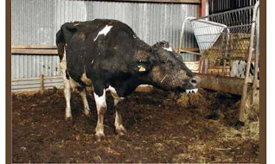
The most consistent signs in cattle are tachypnea and coughing. Initially, rapid, shallow breathing is accompanied by a cough that is exacerbated by exercise. Respiratory difficulty may ensue, and heavily infected animals stand with their heads stretched forward and mouths open and drool. The animals become anorectic and rapidly lose condition. Lung sounds are particularly prominent at the bronchial bifurcation. In adult dairy cattle, milk yield drops severely, and abnormal lung sounds are heard over the caudal lobes. The reinfection phenomenon in adult dairy cattle is usually seen in the fall; although less severe than in initial infections, the signs are widespread coughing and tachypnea and a marked drop in milk yield.
Diagnosis:
Diagnosis is based on CLINICAL SIGN, EPIDEMIOLOGY, presence of first-stage larvae in feces, and necropsy of animals in the same herd or flock. BRONCHOSCOPY and RADIOGRAPHY may be helpful. LARVAE are not found in the feces of animals in the prepatent or postpatent phases and usually not in the reinfection phenomenon (D viviparus). ELISA tests are available in some countries. The test is mainly of use in detecting cattle that have not been exposed, rather than as a differential diagnostic tool in acute respiratory disease. In the early stages of an outbreak, larvae may be few in number. First-stage larvae or larvated eggs can be recovered using most fecal flotation techniques with the appropriate salt solutions; however, larvae will crenate if allowed to sit for a long time on the slide before examination, making identification difficult.
Control:
Lungworm infections in herds or flocks are controlled primarily by vaccination or anthelmintics. Oral vaccines are available in Europe for D viviparus (northeastern areas) and D filaria (southeast). Two doses of irradiated infective larvae are given 4 wk apart, with the second dose given at least 2 wk before the start of grazing or exposure to probable infection. Used properly, they prevent clinical disease, but some vaccinated animals may become mildly infected to the extent that larvae are excreted and perpetuate further infection.
(Verminous bronchitis, Verminous pneumonia)
By Lora R. Ballweber, MS, DVM, College of Veterinary Medicine and Biomedical Sciences, Colorado State University
(The most characteristic clinical sign of lungworm infection is widespread coughing within a herd. Mortality occurs in heavy infections. Although dairy or dairy-cross calves are most commonly affected with lungworm, #Autumn- born single-suckled beef calves are just as susceptible when turned out to grass in early #summer.
